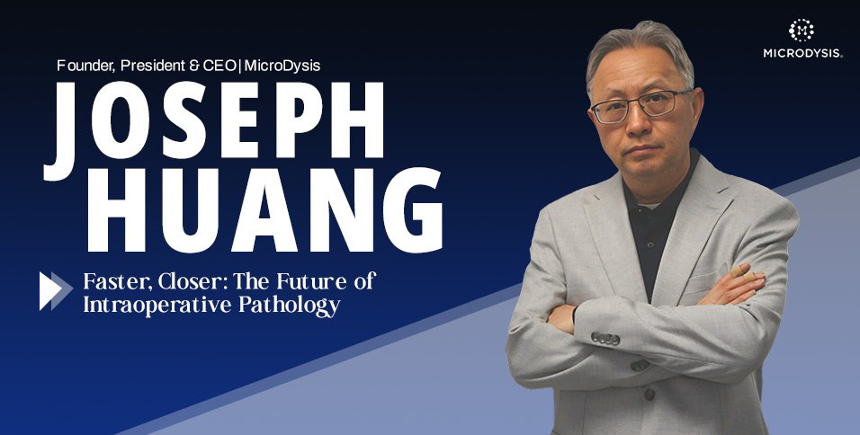
By Joseph Huang, Founder, President & CEO, MicroDysis
A surgeon makes an incision, removes a piece of tissue, and then waits. Somewhere behind the operating room doors, that tissue is being examined so the team can decide what to do next. Is the margin clear? Does the lymph node carry disease? Should more tissue come out? Intraoperative pathology exists precisely because those questions cannot always wait for a later answer. In that pause, minutes matter more than almost anywhere else in medicine.
For more than two decades, I have thought about that pause and the gap between removing tissue and understanding it. As Founder, President, and CEO of MicroDysis, I have focused on how that gap can affect surgical decision-making. My approach is not simply about building a faster machine. It is about rethinking where pathology happens, how it happens, and how close it can get to the moment a decision is actually made.
This is the challenge that has shaped MicroDysis’ work in developing faster, more controlled, and more clinically practical pathology workflows.
The Clock That Never Stops in the Operating Room
Time in surgery is not just a scheduling concern. It is often woven into the clinical decision itself. When a surgeon removes tissue and waits for pathological information, the entire operating room is suspended between action and uncertainty. The faster that tissue can move through preparation, staining, review, and interpretation, the more confidently the surgical team can move forward.
One question has guided much of my work: “How can engineering serve a real clinical decision at the moment that decision matters?” That question has stayed with me since MicroDysis was founded in 2003, long before decentralized diagnostics became an industry buzzword.
Pathology has always been foundational to medicine. It informs diagnosis, staging, and treatment selection, and in cancer care especially, tissue remains one of the most reliable sources of truth. But the traditional pathology model was built around centralized laboratories and carefully sequenced manual steps, a system designed for thoroughness rather than the urgency of an operating room where a decision may need to be made in minutes.
Where Frozen Section Pathology Falls Short
For many years, frozen section pathology has helped surgeons obtain rapid answers during procedures. It has served medicine well, but it also has real limitations. The process is labor-intensive, interpretation can be difficult, and the information it provides may be narrower than what is possible with more advanced staining and molecularly informed methods.
The problem, in other words, is not that any single step is too slow. It is that the whole pathway, from tissue removal to a usable answer, was never designed with the surgical clock in mind. What we need instead is a workflow-compatible system that preserves diagnostic quality while reducing delay, manual handling, and the physical distance between the sample and the decision it is meant to inform.
Rethinking the Microenvironment Where Staining Happens
This is where my engineering perspective meets a clinical workflow problem. At MicroDysis, our long-term focus has been developing the Elim autostainer platform, a system designed around a simple but significant idea: rethink the microenvironment in which tissue staining actually occurs.
The Elim platform is being developed as a microfluidic tissue-staining system designed to support faster, more controlled H&E and immunohistochemistry workflows.
Conventional staining relies on larger reagent volumes, open-slide handling, and a series of manual steps performed on centralized instruments. A more controlled, microfluidic approach can instead manage reagent delivery, incubation, temperature, mixing, washing, and timing directly on the tissue surface itself. This is not simply about making equipment smaller. It is about gaining more precise control over every part of the process.
When that environment is better controlled, staining can become faster, more consistent, and more efficient, using less reagent and producing less variability from one sample to the next. Both standard staining methods and immunohistochemistry may become more suitable for near-OR or point-of-care pathology workflows, particularly in the setting where speed is scarce: the operating room.
Why Intraoperative Pathology Must Be Faster and Trusted
It would be easy to treat speed as the whole story, but I do not. For intraoperative pathology, what matters is not how fast one step in the process runs. What matters is whether the complete pathway, from tissue to decision, can deliver actionable information within the surgical window. A rapid staining method on its own changes little if the surrounding workflow remains slow, manual, or difficult to reproduce.
Healthcare has already moved toward decentralization in other areas. Point-of-care testing has reshaped how clinicians think about blood chemistry, infectious disease screening, glucose monitoring, and cardiac markers. Pathology has been more difficult to decentralize because tissue is not a simple liquid sample. It has structure, architecture, and spatial detail that must be preserved and revealed carefully. That difficulty is precisely why innovation in this area matters.
Our goal is not to replace pathologists or diminish the laboratory’s role. It is to extend the reach of pathology, so expertise can be applied at the moment a surgeon needs it rather than only in the hours that follow.
Building the Foundation Before AI Can Help
Much of the conversation around artificial intelligence in pathology centers on image analysis: spotting tumor cells, measuring biomarkers, and assisting with classification. Those are genuinely valuable directions, but I believe an equally important part of the workflow comes before image analysis. AI is only as strong as the consistency of the data feeding it.
If slide preparation varies, if staining is inconsistent, or if imaging conditions shift from sample to sample, AI performance suffers no matter how sophisticated the algorithm is. That means the future of AI-enabled pathology has to begin before any image is ever analyzed. It starts with tissue handling, staining consistency, and standardized image acquisition. A reliable front end is what makes reliable downstream intelligence possible at all.
The Team Behind the Technology
Building a company like this takes more than a working prototype. It takes people who can turn a scientific vision into something investable and sustainable.
Molly Sung, Vice President of Business Development, has helped MicroDysis build relationships and support fundraising through SAFE financing, drawing on her broad connections in the real estate and investment community. Our board members, Hanan Davidowitz and Patric Jones, provide important guidance from the biotech business and legal perspectives, helping strengthen the company’s path from invention toward commercialization.
Moving an invention toward practical use requires more than technology alone. Trust, partnerships, and capital structure have to be built alongside the technology, not after it.
Why This Matters Beyond the Operating Room
The implications of faster, more automated tissue staining reach well past a single procedure. Smaller hospitals, ambulatory surgical centers, remote clinical settings, and research environments that lack large centralized infrastructure could all benefit from systems that bring timely pathological insight closer to the point of care.
As healthcare systems everywhere are asked to do more with fewer resources, reducing manual steps and standardizing workflows is not a luxury. It is becoming part of what practical, sustainable medicine looks like.
At the same time, ambition must be tempered with discipline: “Diagnostic technologies must earn trust through validation, reproducibility, clinical comparison, and regulatory discipline. Speed alone is not enough.”
What I envision instead is a system in which central laboratories are increasingly supported by near-OR and point-of-care pathology technologies that deliver specific, time-sensitive answers rapidly, with pathologists’ expertise extended through better preparation systems, digital imaging, and eventually AI-assisted tools, even when they cannot be physically present in every operating room.
Closing the Distance Between Tissue and Decision
For me, the future of intraoperative pathology is not simply about faster machines or more powerful algorithms. It is about bringing clearer diagnostic insight to the right people at the right moment. In surgery, that moment can be measured in minutes.
Closing the distance between tissue and decision is therefore not just a technology challenge; it is a leadership responsibility. It requires scientific discipline, clinical awareness, and the patience to build systems that can earn trust.
The path forward will not be defined by speed alone. It will be defined by whether faster, more consistent pathology can become genuinely useful at the point where surgical decisions are being made.
The real measure of progress in intraoperative pathology will be whether technology can shorten the distance between what clinicians need to know and when they need to know it.