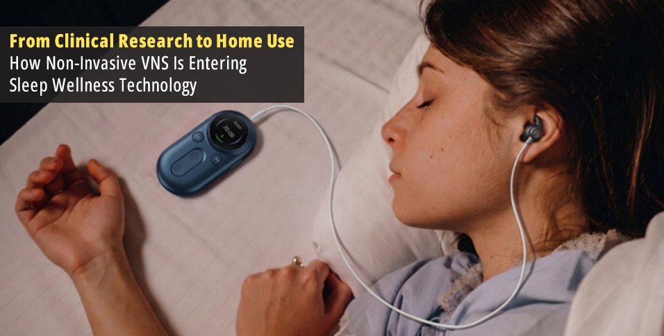
A device that sends small electrical pulses through the ear is a long way from the implanted systems doctors have used for vagus nerve stimulation for decades. Yet both approaches belong to a growing field of neuromodulation research, and the ear is becoming an increasingly practical place to study how electrical stimulation interacts with the nervous system.
Part of the interest comes from sleep research. Scientists have begun testing whether transcutaneous auricular vagus nerve stimulation, or taVNS, can help people with chronic insomnia. At the same time, companies are developing smaller devices that can be worn during an evening relaxation routine without surgery or a visit to a clinic.
Bringing this technology home involves more than shrinking the hardware. Researchers first need to understand which parts of the ear can carry relevant signals, how those signals reach the brain, and whether stimulation produces meaningful changes in sleep. Device makers then face another set of challenges: keeping electrodes in the right place, delivering consistent pulses, and making the equipment comfortable enough for regular use.
Why the Ear Became a Target for Vagus Nerve Stimulation
The vagus nerve runs from the brainstem into the neck, chest, and abdomen, carrying information between the brain and several internal organs. Most of its fibers are sensory, sending information toward the brain rather than carrying instructions away from it. That makes the vagus nerve more than a pathway for slowing the heart or helping the body relax.
Traditional implanted VNS uses an electrode placed around the cervical vagus nerve in the neck. Auricular VNS takes advantage of a smaller sensory branch that supplies parts of the outer ear. Electrical pulses applied through the skin can stimulate sensory fibers in these regions without placing an electrode inside the body.
The location matters because the outer ear is not supplied by one nerve alone. In a dissection study involving 14 ears from seven cadavers, Peuker and Filler (2002) mapped a mixture of vagal and non-vagal nerve supply. The cymba conchae, the recessed area in the upper part of the ear’s bowl, was identified as an important region of vagal innervation. Other areas, including the cavum conchae below it, may receive input from multiple sensory nerves.
This anatomical work helped give researchers a more specific target than simply placing electrodes somewhere on the ear. For wearable developers, it also created an engineering question: how can a small device maintain reliable contact with an anatomically relevant region when ear shapes vary from person to person?
That question is now part of the design of ear-worn vagus nerve stimulation technology, including the non-invasive devices offered by ZenoWell. But reaching the correct area is only the first step. Researchers still need to show what happens after stimulation begins.
How Researchers Test Whether Ear Stimulation Reaches the Brain
One way to investigate ear stimulation is to watch how the brain responds. In 2015, Frangos and colleagues studied 12 healthy adults using functional magnetic resonance imaging (fMRI). They applied electrical stimulation to the left cymba conchae and compared the brain response with stimulation at the earlobe, an area commonly used as a control in this kind of research.
The cymba stimulation produced activity changes in regions associated with vagal pathways, including the nucleus tractus solitarius (NTS). Located in the brainstem, the NTS helps process sensory information arriving from internal organs and connects with wider networks involved in autonomic regulation and arousal.
The researchers did not measure whether participants slept better afterward. Their experiment addressed an earlier question: could stimulation at the outer ear engage brain regions associated with vagal signaling?
Other studies have examined the electrical responses produced by stimulation at different ear locations. These include vagal somatosensory evoked potential studies, often shortened to VSEP studies. Researchers record electrical signals following stimulation and compare how the recorded responses change with electrode placement and other conditions.
García de Gurtubay and colleagues (2021) examined different auricular stimulation sites and electrode configurations. Their findings provided additional reasons to investigate combined coverage of the cymba and cavum conchae, although the interpretation of evoked potentials as specific measures of vagal activation remains an area of scientific discussion.
The distinction between these research methods is worth understanding:
| Research method | What researchers examine | What it cannot establish by itself |
| Anatomical dissection | Which nerves supply different parts of the ear | Whether stimulation improves sleep |
| Functional MRI | Changes in brain activity during stimulation | Whether those changes produce a clinical benefit |
| Evoked potential recordings | Electrical responses associated with stimulation | Whether one electrode design delivers better health outcomes |
| Randomized clinical trials | Differences in outcomes between active and control interventions | Whether the results apply to every consumer device |
Together, these studies help explain why researchers focus on particular ear regions. They do not show that all electrodes placed near those regions work equally well, or that a stronger recorded signal necessarily leads to better sleep.
There is also an important difference between ear and neck stimulation. Cervical devices deliver electrical pulses through tissues around the neck, while auricular devices work through superficial sensory regions of the ear. Their electrode placement, contact requirements, and stimulation characteristics are not interchangeable.
Readers considering either approach can explore the practical differences in this guide to ear versus neck vagus nerve stimulation, including how device placement and intended use affect the choice.
What the Sleep Trials Have Found So Far
Knowing that stimulation can produce a brain response is useful, but it does not answer whether someone will sleep better. For that, researchers need clinical trials with measurable sleep outcomes and appropriate comparison groups.
A randomized clinical trial published in JAMA Network Open studied 72 adults with chronic insomnia disorder (Zhang et al., 2024). Participants received either active taVNS or sham stimulation for eight weeks, with sessions lasting 30 minutes twice daily on five days each week. Researchers measured changes using the Pittsburgh Sleep Quality Index (PSQI), a questionnaire covering several aspects of sleep over the preceding month.
Both groups reported improvements, but the active-stimulation group improved more. At eight weeks, the adjusted average PSQI score had fallen by 8.2 points in the active group compared with 3.9 points in the sham group. The between-group difference was approximately 4.2 points, exceeding the study’s predefined threshold for a clinically meaningful change. The study also followed participants for another 12 weeks, with the reported group differences maintained during follow-up.
These are meaningful results from a controlled trial, not simply a theoretical claim about the vagus nerve. However, the study was conducted at one clinical center, involved patients diagnosed with chronic insomnia, and used a specific stimulation schedule. It did not establish whether a shorter consumer wellness session would produce similar results.
A 2025 systematic review and meta-analysis by de Oliveira and colleagues brought together six studies involving 336 patients with insomnia. The pooled results favored taVNS for sleep quality and insomnia severity, but the authors rated the certainty of evidence as low for sleep quality and very low for insomnia severity.
That leaves researchers with several questions. Different studies have used different stimulation parameters and treatment schedules, making it difficult to identify a single protocol that works best. Many outcomes also rely on questionnaires, so further work using objective sleep measurements and longer follow-up would help clarify the findings.
For consumers, the distinction is between a technique that has shown encouraging results under studied clinical conditions and a particular home device whose sleep benefits may not have been independently established.
Why Electrode Design Matters Outside the Laboratory
In a research setting, trained staff can check electrode position, adjust stimulation according to the protocol, and monitor what participants feel. At home, the person using the device usually has to manage those steps independently.
Electrical contact is one of the first challenges. The curved surface of the ear makes it difficult for electrodes to sit in exactly the same way on everyone. Skin moisture, pressure, electrode size, and placement can all affect electrical impedance, the resistance encountered by the current as it passes through the contact area.
If contact changes during a session, the sensation may change as well. An electrode may feel stronger against one part of the ear than another, or the sensation may fade as the skin dries or the contact shifts. That does not necessarily mean the device has stopped working, and turning up the intensity is not always the appropriate response.
ZenoWell’s product training materials describe an ear-contact design informed by anatomical research on the cymba and cavum conchae, with attention to stable contact and comfortable positioning. Its Sleep mode provides a useful example of how a manufacturer translates a stimulation concept into a defined user session.
According to the training materials, the mode uses alternating 20 Hz and 4 Hz stimulation, a 200-microsecond pulse width, and a 20-minute session. The programmed cycle consists of seven seconds at 20 Hz followed by three seconds at 4 Hz. Users can select this mode directly on the device.
Those settings describe ZenoWell’s product design. They should not be confused with the protocol used in the Zhang et al. clinical trial or presented as clinically established optimal settings for insomnia.
The company’s instructions also describe checking that both electrode contacts rest against clean skin, beginning at a low intensity, and keeping the sensation noticeable but comfortable. The device is intended to be removed after the timed session rather than worn overnight.
These details matter because a home-use product has to work in an ordinary environment, not just under research supervision. Someone preparing for bed needs to understand how to position the device, what a normal sensation may feel like, and when discomfort means they should stop.
What Still Needs to Be Established for Home Use
Non-invasive stimulation removes the need for an implanted electrode, but it does not remove every safety consideration. A 2022 systematic review and meta-analysis by Kim and colleagues examined adverse events reported across 177 taVNS studies involving 6,322 participants. Reported problems were generally mild and temporary, including ear discomfort, headache, and tingling. However, adverse-event reporting was incomplete in many studies, making the available evidence less comprehensive than the number of participants might suggest.
For home-use devices, manufacturers need clear precautions and instructions that people can follow without a clinician present. ZenoWell’s training materials advise against using its device while driving, bathing, or operating machinery, and against placing electrodes over wounded or inflamed skin. They also identify implanted electronic medical devices, seizure history, pregnancy, and certain cardiac conditions as circumstances requiring avoidance or prior medical assessment.
The research also leaves a separate question about effectiveness. Larger trials may help researchers understand which patients respond, which stimulation schedules are most useful, and whether improvements persist. Product-specific testing is needed before those findings can be attributed to an individual consumer device.
There is a practical opportunity here for wearable technology. Researchers have established reasons to investigate the ear as a stimulation site, developed ways to measure neural responses, and reported encouraging clinical sleep results. Device makers are working on the less visible problems that determine whether the technology can be used outside a clinic: reliable skin contact, tolerable stimulation, straightforward controls, and clear safety instructions.
Non-invasive VNS is becoming more accessible, but accessibility and proven sleep benefits are separate achievements. As ear-worn devices move into everyday wellness, the next important advances will come from bringing better home-use design together with stronger evidence for the devices people actually buy.