The search for durable aesthetic corrections shifted the focus of regenerative science toward bioresorbable polymers. Temporary soft tissue augmentation used to rely heavily on simple volume replacement; substances filled space, held water, and gradually disappeared without leaving much behind. Polycaprolactone microspheres changed that dynamic completely. Injectable materials can now act as scaffold architectures, directing local cell populations to produce fresh structural proteins as the synthetic matrix breaks down.
Scientists originally evaluated polycaprolactone, or PCL, for surgical sutures and drug delivery systems because the body tolerates it exceptionally well. When processed into perfectly smooth spherical particles and suspended in a hydrogel matrix, its behavior inside living tissue becomes remarkably predictable. The physical presence of these tiny spheres triggers a targeted biological reaction that outlasts the gel that delivered them.
Mechanism of Action and Matrix Formulation
To grasp how a microsphere-based dermal filler functions, you have to look closely at the primary ingredients. The formulation consists of two separate components operating on different timelines. The visible phase starts with a carboxymethylcellulose carrier gel. This viscous gel provides immediate volume correction right after placement, filling structural voids and smoothing out deep folds.
Carboxymethylcellulose does not stay in the tissue forever. The body breaks down and absorbs this gel carrier over a period of several weeks. If the product contained only hydrogel, the initial cosmetic effect would collapse quickly. Suspended evenly throughout that gel, however, are smooth polycaprolactone microspheres making up roughly thirty percent of the total volume. These particles range between 25 and 50 micrometers in diameter. That specific size window matters immensely: smaller particles risk immediate phagocytosis by macrophages, which causes rapid clearance and potential inflammation, while larger or irregularly shaped particles can provoke foreign body granulomas.
The aesthetic market features specific commercially available options engineered around this exact dual-action concept. Ellanse stands out as a primary example of a collagen stimulating filler built on PCL technology. Medical professionals select Ellanse when patients require both instant volumetric lifting and long-lasting structural support. The formulation relies on smooth, perfectly spherical PCL structures to trigger natural tissue regeneration as the primary gel dissipates over time. Clinics often look to certified distributors to shop Ellanse online for clinic use when maintaining inventory for bio-stimulatory procedures. Utilizing Ellanse allows practitioners to offer tailored treatment plans based on predictable biological longevity rather than simple volume replacement.
Polymer Physics and Hydrolytic Degradation Kinetics
How does polycaprolactone actually break down inside human tissue? The process differs significantly from other biodegradable materials like polylactic acid or cross-linked hyaluronic acid. PCL undergoes a clean bulk hydrolysis process. Water molecules from surrounding biological fluids penetrate the amorphous regions of the polymer chains, breaking the ester bonds that hold the molecular structure together.
During the initial phase of dermal implant biodegradation, the molecular weight of the polymer drops continuously, yet the physical size and overall shape of the individual microspheres remain intact. The structural integrity of each sphere does not collapse right away. This predictable, slow loss of molecular weight without immediate structural fragmentation explains why clinical volume remains constant for long periods.
The polymer chain breakdown happens in a two-stage pattern:
- Bulk degradation stage: Ester bonds cleave throughout the entire sphere without changing the exterior dimensions or overall mass of the particle.
- Surface erosion stage: Once the polymer chains reach a critical lower molecular weight threshold, mass loss finally begins; the fragments convert into caproic acid, which the body metabolizes through normal metabolic pathways into carbon dioxide and water.
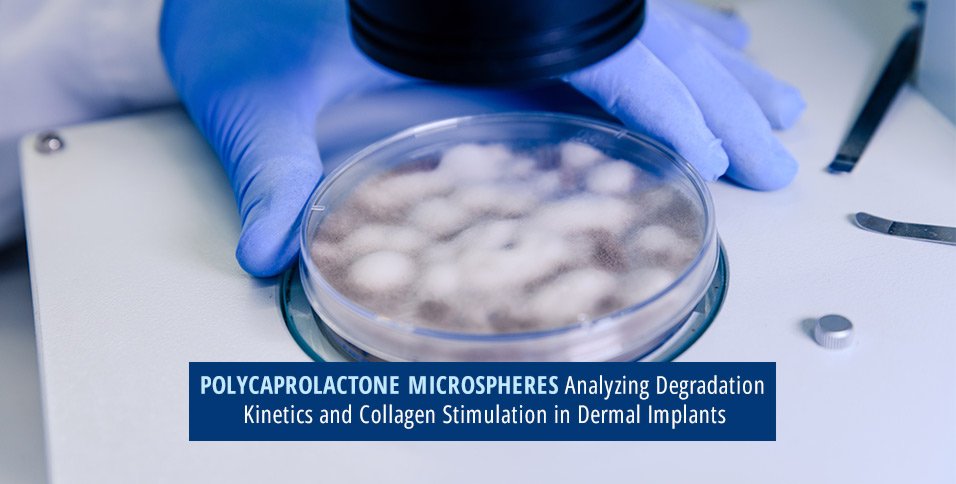
A 2017 study examining PCL biodegradation mechanics highlighted that the degree of polymer crystallinity and initial molecular weight directly dictate the implant degradation timeline. By manipulating these chemical properties during manufacturing, engineers can predetermine how long the microspheres resist mass loss, yielding predictable degradation kinetics across different product variants.
Neocollagenesis and Structural Tissue Remodeling
While PCL microsphere breakdown proceeds quietly in the background, the tissue response around those spheres drives the secondary phase of volume maintenance. The body recognizes the microspheres as mild foreign objects, initiating a controlled, non-inflammatory tissue reaction. Fibroblasts migrate toward the site, anchoring themselves to the smooth surface of the polycaprolactone structures.
This cellular activity triggers robust collagenesis PCL filler responses. The primary structural protein generated during this process is collagen. Early after placement, histology reveals the formation of Type III collagen, the flexible type associated with wound healing and initial matrix deposition. Over subsequent months, local tissue remodeling replaces Type III collagen with dense, highly organized Type I collagen, the main structural protein responsible for skin firmness and mechanical resistance.
Research evaluating histological samples demonstrates that long-term collagen production creates a natural extracellular matrix surrounding each sphere. The new tissue holds the space as the artificial microspheres slowly lose structural integrity. Instead of leaving empty tissue pockets upon complete degradation, the original synthetic volume gets replaced almost entirely by the host’s own structural framework.
Clinical Safety and Comparative Advantages
Safety profiles in biostimulatory materials rely heavily on particle surface morphology. Sharp edges or irregular surfaces on microspheres excite immune cells, causing persistent inflammation, giant cell reactions, and visible nodules under the skin. Polycaprolactone microspheres feature smooth, spherical surfaces, minimizing mechanical friction against surrounding tissue structures.
When you compare a gradual biodegradation implant based on PCL to traditional hyaluronic acid gels, the fundamental difference lies in longevity and material properties. Hyaluronic acid holds water mechanically; it expands tissue by binding fluid molecules, degrading gradually via enzymatic activity. PCL operates as a true biological scaffold. The carrier gel handles short-term cosmetic needs, but the long-term volume stems from genuine autologous tissue growth.
Risk profiles remain low when qualified clinicians place the material using correct anatomical depth and technique. Because PCL cannot be dissolved instantly with an enzyme like hyaluronidase, precise placement in deeper dermal or sub-dermal layers remains critical. Deep placement ensures the newly formed collagen network sits beneath the superficial dermis, providing subtle structural support without creating visible surface irregularities.
The development of PCL microsphere systems represents a major step forward in soft tissue augmentation science. Integrating controllable chemical degradation with predictable tissue regeneration bridges the gap between short-acting fillers and permanent, non-resorbable implants. As researchers continue refining polymer chemistry and processing techniques, the ability to fine-tune both the rate of hydrolytic degradation and the volume of generated tissue will only become more precise.