According to the American Dental Association’s 2023 oral health surveillance data, periodontal disease affects nearly half of adults over 30 — and among those cases, the progression to bone loss often happens silently, with many patients unaware until structural damage becomes irreversible. As dental imaging technology advances and preventive care shifts toward early intervention, the ability to catch jaw bone deterioration in its earliest stages has become a critical factor in preserving both function and facial structure.
The stakes extend far beyond oral health. When jaw bone loss progresses unchecked, it triggers a cascade of problems: teeth become loose and may fall out, facial features begin to collapse inward, and even the simplest activities like eating and speaking become compromised. For the thousands of Americans facing this progression each year, recognizing the subtle warning signs can mean the difference between a minor intervention and major reconstructive surgery.
What makes early detection particularly challenging is that initial bone loss often presents through seemingly minor symptoms — slight gum changes, occasional discomfort, or barely perceptible shifts in how teeth feel. Understanding these early indicators, their underlying causes, and the treatment pathways available provides the foundation for making informed decisions about oral health before irreversible damage occurs.
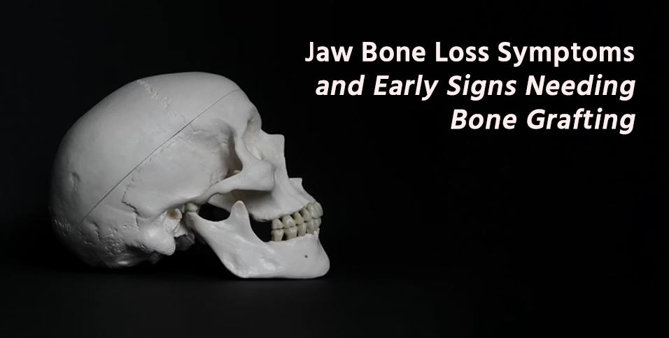
What Early Symptoms Indicate Jaw Bone Loss
The earliest signs of jaw bone deterioration often masquerade as minor dental annoyances, making them easy to dismiss until the underlying problem becomes severe. Gum recession represents one of the most reliable early indicators — as bone support diminishes, the gums gradually pull away from the teeth, exposing more of the tooth surface than normal. This recession typically happens slowly, which explains why many people don’t notice it until someone points it out or they compare current photos to older ones.
Tooth mobility provides another critical warning sign, though it requires attention to subtle changes. Healthy teeth should feel completely stable when you press on them with your tongue or finger. The first hint of bone loss often appears as a barely perceptible “give” when pressure is applied, or teeth that feel slightly different when chewing firm foods. This mobility occurs because the bone that normally anchors tooth roots firmly in place begins to dissolve away, reducing the structural support system.
Pain patterns associated with early bone loss differ from typical toothaches. Rather than sharp, localized pain, bone loss often produces a dull, persistent ache in the jaw area, particularly when waking up in the morning. This discomfort may intensify when chewing tougher foods or may present as a general soreness that seems to move around the mouth rather than staying in one specific tooth.
Functional changes provide additional clues that bone support is compromised. Many people notice their bite feels “different” — teeth that once fit together perfectly now seem slightly misaligned, or there’s a new gap where food gets caught. Denture wearers often experience the first signs of bone loss through changes in how their appliances fit; dentures that once felt secure may become loose or develop sore spots as the underlying bone structure changes shape.
The challenge with these early symptoms lies in their subtlety and gradual progression. Unlike a broken tooth or severe cavity that demands immediate attention, bone loss symptoms develop over months or years, allowing people to adapt unconsciously to the changes until the damage becomes extensive.
What Causes and Risk Factors Contribute to Jaw Bone Loss
Common Biological and Systemic Causes
Periodontal disease stands as the leading cause of jaw bone deterioration, accounting for the majority of bone loss cases in adults. The bacterial infections that characterize gum disease produce toxins that trigger the body’s immune response, but this protective reaction inadvertently destroys the very bone tissue it’s trying to protect. As bacteria colonize the spaces between teeth and gums, the resulting inflammation creates an environment where bone-dissolving cells outnumber bone-building cells, leading to progressive deterioration.
Tooth extraction initiates an immediate bone loss process that many people don’t anticipate. Within the first year after extraction, the bone that once supported that tooth can lose up to 25% of its width, as the body recognizes the bone is no longer needed for structural support. This remodeling process continues indefinitely, which explains why people who’ve had teeth removed years earlier often struggle with ill-fitting dentures or notice changes in their facial profile.
Osteoporosis and systemic diseases accelerate bone loss throughout the body, including the jaw. Conditions like diabetes compromise the body’s ability to fight infections and heal properly, making periodontal disease more likely to progress to bone loss. Autoimmune disorders can trigger inflammation that affects bone metabolism, while certain medications — particularly those used for osteoporosis treatment or cancer therapy — can interfere with normal bone renewal processes in unexpected ways.
Lifestyle and Demographic Risk Factors
Age-related factors compound other risk elements, as bone density naturally decreases after age 30. However, chronological age matters less than cumulative exposure to risk factors — a 40-year-old with untreated gum disease may experience more bone loss than a healthy 70-year-old with excellent oral hygiene. The key lies in understanding that aging doesn’t automatically cause bone loss, but it does reduce the body’s ability to repair damage quickly.
Smoking creates a particularly destructive environment for jaw bone health by reducing blood flow to the gums and bone tissue. Nicotine constricts blood vessels, limiting the nutrients and immune cells that reach the area, while the chemicals in tobacco smoke interfere with the cellular processes that rebuild bone. Smokers experience bone loss at rates up to three times higher than non-smokers, and the damage often progresses faster and responds more poorly to treatment.
Nutritional deficiencies — particularly inadequate calcium, vitamin D, and phosphorus — compromise the body’s ability to maintain bone density. However, nutrition’s role extends beyond basic minerals; protein intake affects bone regeneration, while excessive alcohol consumption interferes with the hormonal systems that regulate bone metabolism. Women face additional risks during menopause, when declining estrogen levels accelerate bone loss throughout the body, including the jaw.
How Jaw Bone Loss Affects Facial and Oral Structure
The architectural role that jaw bone plays in facial structure becomes apparent only when that support begins to fail. As bone volume decreases, the facial profile undergoes gradual but dramatic changes — the lower third of the face begins to collapse inward, creating a sunken appearance around the mouth and cheeks. This transformation happens slowly enough that family members often don’t notice until they compare recent photos to images taken several years earlier, revealing changes that seemed to occur overnight but actually developed over months or years.
Denture stability deteriorates as the underlying bone shrinks, creating a cycle where poorly fitting dentures accelerate further bone loss. When dentures don’t fit properly, they create pressure points that can damage the remaining bone tissue, while the lack of stimulation from tooth roots causes the bone to remodel away. Many long-term denture wearers eventually reach a point where traditional dentures become virtually impossible to wear comfortably, regardless of adjustments or adhesives.
The structural changes extend beyond aesthetics to affect basic oral functions. Tooth stability throughout the mouth becomes compromised as adjacent teeth lose their anchor points and begin to shift. Even teeth that aren’t directly affected by localized bone loss may become loose as the overall architecture of the mouth changes. This shifting can create new spaces where food accumulates, establishing conditions that promote further periodontal problems and additional bone loss.
Speech patterns often change as teeth become mobile or as facial structure shifts. Sounds that require precise tongue-to-tooth contact — like “th,” “s,” and “f” sounds — may become difficult to pronounce clearly. Some people develop a slight lisp or find that their dentures click when speaking, particularly during phone conversations when they’re concentrating more on word formation than usual.
The psychological impact of these changes shouldn’t be underestimated. Many people report avoiding social situations, covering their mouth when laughing, or choosing softer foods not just for comfort but to avoid embarrassment. When considering the significant role that bone grafting in Green River plays in restoring both function and confidence, early intervention becomes crucial for preventing these life-altering consequences. The progression from minor gum recession to major facial structural changes typically spans several years, providing multiple opportunities for intervention if the warning signs are recognized and addressed promptly.
How Is Jaw Bone Loss Diagnosed Early
Early detection of jaw bone loss relies on a combination of clinical examination techniques and advanced imaging that can reveal changes before they become visible to the naked eye. During routine dental examinations, practitioners use specialized probes to measure the depth of spaces between teeth and gums — healthy pockets typically measure 1-3 millimeters, while measurements of 4 millimeters or greater suggest bone loss may be occurring. This probing also reveals bleeding patterns that indicate active inflammation and bone destruction.
Digital X-rays provide the most reliable method for documenting bone levels and tracking changes over time. Unlike older film X-rays that required significant bone loss before changes became apparent, modern digital imaging can detect bone density changes as small as 5-10%. Practitioners typically take a series of images from different angles to create a comprehensive map of bone levels around each tooth, establishing baseline measurements for future comparison.
Advanced diagnostic tools have revolutionized early detection capabilities. Cone beam CT scans create three-dimensional images that reveal bone thickness and density patterns impossible to see on traditional X-rays. These detailed images allow practitioners to identify areas where bone loss is just beginning and to plan treatment approaches before structural damage becomes extensive. Some practices now use specialized imaging software that compares current bone levels to previous scans, highlighting even subtle changes that might otherwise be overlooked.
The timing of diagnostic evaluations plays a crucial role in early detection. Rather than waiting for symptoms to develop, regular monitoring every 6-12 months allows practitioners to track gradual changes and intervene at the optimal moment. This proactive approach proves particularly valuable for patients with risk factors like diabetes, smoking history, or family history of periodontal disease, where bone loss may progress more rapidly than average.
What Treatment Options Are Available for Jaw Bone Loss
Bone Grafting and Surgical Interventions
Bone grafting procedures represent the most direct approach to rebuilding lost jaw bone tissue, using materials that stimulate new bone growth in areas where natural bone has been lost. The process typically involves placing grafting material — which may come from the patient’s own body, processed donor bone, or synthetic substitutes — into the deficient area and allowing several months for integration. Modern grafting materials have success rates exceeding 90% when performed under optimal conditions, making this a highly predictable treatment option.
Guided bone regeneration enhances the grafting process by using specialized membranes that create a protected space for new bone to grow while keeping soft tissue from interfering with the healing process. This technique proves particularly effective in areas where bone loss has created complex defects that simple grafting alone cannot address. The combination of grafting materials and barrier membranes allows practitioners to rebuild bone architecture that closely matches the original anatomy.
Dental implant integration often works synergistically with bone grafting procedures, as implants themselves stimulate ongoing bone maintenance through the mechanical forces they transmit during chewing. In cases where bone loss has progressed significantly, the grafting procedure may be performed months before implant placement, allowing time for the new bone to mature and develop the density necessary to support long-term implant success.
Non-Surgical Therapies and Preventative Measures
Medications and therapeutic interventions can slow or halt the progression of bone loss, particularly when combined with improved oral hygiene practices. Prescription antimicrobial rinses help control the bacterial populations that trigger bone-destroying inflammation, while certain medications used to treat osteoporosis have shown promise in preserving jaw bone density. However, these approaches work best when bone loss is detected early, before extensive damage has occurred.
Laser therapy and regenerative techniques offer promising alternatives to traditional surgical approaches, using focused light energy to eliminate bacteria and stimulate healing in periodontal pockets. Some practitioners combine laser treatment with growth factors or other biological materials that encourage natural bone regeneration. While these techniques may not replace the need for grafting in advanced cases, they can be highly effective for early-stage bone loss or as adjunctive treatments to enhance healing after surgical procedures.
The most effective approach often involves recognizing that bone loss rarely results from a single cause, requiring treatment plans that address multiple contributing factors simultaneously. For patients facing early-stage bone loss, the window for non-surgical intervention remains open, but that window typically closes as the condition progresses, making early detection and prompt treatment crucial for preserving both oral health and the structural integrity of facial features.