
Most people have occasionally noticed an eye floater drifting through their vision at some point in their lives. In this digital era, up to 76% of people experience these eye floaters. But what happens when that fleeting speck becomes persistent, like a shadow in vision, a dark curtain over vision, a gray patch, different from the normal sight? This change is not something to scroll past.
In some cases, a sudden appearance of a shadow in vision may indicate a retinal tear, retinal detachment, bleeding inside the eye, or a blockage of the retinal blood vessels, all of which could lead to vision loss if ignored for even a few hours.
Because the causes of vision shadow can vary widely, it is important to understand what the symptom means and when it should be considered an emergency. Paying attention to accompanying symptoms such as flashes of light, new floaters, blurred vision, or loss of peripheral vision can help identify potentially serious problems early.
In this article, we’ll explore the common causes of a shadow in vision, associated warning signs, risk factors, available treatment options, and the situations that require urgent evaluation by an eye care professional.
What Does a Shadow in Vision Mean?
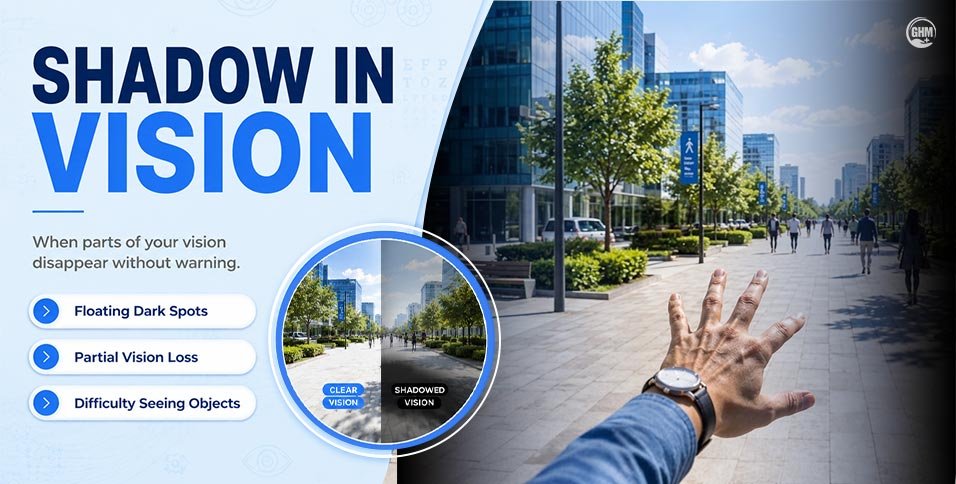
A shadow in vision refers to any area of the visual field that appears darker, grayer, or simply absent. People describe shadow over vision in different ways:
- A dark or gray patch in vision in one part of the sight
- A veil or curtain across vision, as if something is being drawn across the eye
- A missing or blind spot in vision, especially toward the edges
- A peripheral vision shadow that shifts when they move their eyes
The location of the shadow in vision matters. A shadow in the outer (peripheral) part of vision often points to retinal issues, while a central shadow may suggest problems involving the macula or the blood vessels supplying it.
Consequently, it is equally important to know whether the shadow is moving, expanding, or fixed. A shadow that stays in one place after a few days is less alarming than one that grows or shifts rapidly, which demands urgent evaluation.
Common Symptoms Accompanying Shadow in Vision
A shadow in vision rarely appears in isolation. Watch for these accompanying symptoms, because when several occur together, they often signal a more urgent retinal problem:
- Flashes of light, sudden streaks or sparks, particularly in dim lighting
- A sudden surge in eye floaters, more than usual, or entirely new
- Blurred vision, even after blinking
- Wavy or distorted vision, where straight lines appear bent
- Loss of side vision or a narrowing of the visual field
- Reduced contrast sensitivity, difficulty distinguishing shades
- A sudden, noticeable drop in overall visual clarity
If some experience flashes, floaters, and a shadow in vision simultaneously, treat it as an emergency.
What Causes a Shadow in Vision?
Several conditions can cast a shadow across vision. Understanding each one helps judge how urgently it needs care.
1. Retinal Detachment
Retinal detachment is the most serious cause of a shadow in vision. Retinal detachment requires urgent surgical repair to prevent permanent vision loss.
Retinal detachment occurs when the retina separates from the layer of tissue beneath it, cutting off its blood supply. A detached retina can cause a curtain coming down over one side of the visual field. It is a shadow that often starts in peripheral vision and advances inward, indicating a medical emergency.
2. Retinal Tear
An emerging shadow at the edge of vision, a sudden increase in floaters, and flashes of light are symptoms of retinal tear. A retinal tear occurs when the vitreous gel pulls away from the retina forcefully enough to create a small break. Fluid can seep through this break, causing shadows in vision to appear.
Retinal tears are serious but highly treatable when caught early, often with a simple laser procedure performed in the clinic.
3. Eye Floaters and Vitreous Changes
The vitreous is the clear, gel-like substance that fills the inside of the eye. With age, it naturally becomes more liquid and can develop small clumps or strands. These clumps cast shadows on the retina, appearing as floaters or faint gray shapes.
This process, called posterior vitreous detachment (PVD), is very common and usually harmless. However, it can occasionally trigger a retinal tear, so new or worsening floaters always signal a dilated eye exam.
4. Vitreous Hemorrhage
When blood leaks into the vitreous cavity, often due to diabetes, eye trauma, or a torn retinal vessel, it can create dark spots, reddish-brown shadows, or a cobweb-like haze. This is called a vitreous hemorrhage.
In people with diabetic retinopathy, new blood vessels on the retina are fragile and prone to bleeding, making vitreous hemorrhage a common complication.
5. Retinal Blood Vessel Blockage
A blockage in the arteries or veins supplying the retina, known as retinal artery occlusion or retinal vein occlusion, can cause sudden, painless loss of vision in part or all of the visual field. The affected area often appears as a shadow or gray-out. This condition is treated as a stroke of the eye and requires immediate attention.
Risk Factors Leading To A Shadow in Vision
Certain groups face a higher risk of experiencing shadow in the eye.
New visual symptoms to monitor if someone notices these risk factors:
- Age-Related Eye Changes: The vitreous gel degrades naturally with age. Adults over 50 are significantly more likely to experience posterior vitreous detachment and related retinal complications.
- High Myopia (Severe Nearsightedness): Highly myopic eyes are physically longer than average, which stretches and thins the retina, making it more susceptible to tears and detachment.
- Diabetic Retinopathy: Poorly controlled blood sugar damages the tiny vessels in the retina over time. Diabetic retinopathy affects an estimated 1 in 3 people with diabetes and is a leading cause of vitreous hemorrhage and shadow-related vision changes.
- Previous Eye Surgery: Cataract surgery and other intraocular procedures can occasionally lead to vitreous changes or retinal traction that increases the chance of a retinal tear or detachment post-operatively.
- Eye Injury or Trauma: A direct blow to the eye or head can cause retinal tears, detachment, or vitreous bleeding, sometimes even days or weeks after the initial injury.
- Family History of Retinal Disease: A family history of retinal detachment, macular degeneration, or other inherited retinal conditions raises the personal risk.
How Eye Doctors Diagnose the Cause of a Shadow in Vision
Early diagnosis is the most important factor in preserving vision. A curtain shadow that moves or grows is highly suggestive of progressive retinal detachment and should never be observed at home. Visiting an eye specialist is an accurate step.
An eye specialist will use one or more of the following tools for vision shadow diagnosis:
- Comprehensive eye examination is the starting point for any eye issue.
- Dilated retinal exam allows the doctor to see the full retina, including its edges.
- Optical Coherence Tomography (OCT) is a non-invasive scan that creates detailed cross-sectional images of retinal layers.
- Retinal imaging is a wide-field photography to map the entire retina.
- Ocular ultrasound (B-scan) is used when the view inside the eye is blocked by bleeding.
- Visual field testing measures the full extent of patient’s peripheral and central vision.
Treatment Options for Shadow in Vision
Treatment for the shadow across vision is determined entirely by the underlying cause.
1. Observation for Benign Floaters:
When symptoms of curtain shadow are mild, stable, and linked to a benign posterior vitreous detachment, the doctor may recommend monitoring with scheduled follow-up visits.
2. Laser Treatment (Laser Photocoagulation)
For retinal tears detected before detachment occurs, laser photocoagulation seals the tear by creating small burn marks around it, preventing fluid from passing underneath. This is typically a quick, in-clinic procedure.
3. Cryopexy
Cryopexy uses controlled freezing to create scar tissue around a retinal break, sealing it securely. It is often used alongside other retinal repair procedures.
4. Vitrectomy
The vitrectomy procedure removes the vitreous gel, which may be causing traction on the retina or harboring blood from a vitreous hemorrhage. It is a microsurgical procedure performed under local or general anesthesia, with an 85% to 90% success rate.
5. Retinal Detachment Surgery
Depending on the type and extent of detachment, surgeons use one or a combination of:
- Scleral buckle, a silicone band placed around the eye to push the wall closer to the detached retina
- Pneumatic retinopexy, a gas bubble injected into the eye to press the retina back into place
- Vitrectomy-based repair combined with gas or silicone oil tamponade
End Note
A shadow in vision can range from a harmless sign of aging to a true ocular emergency. The difference often comes down to how quickly we act.
The shadow in vision caused by benign vitreous changes can be safely monitored, but retinal tears and retinal detachment demand same-day care. When a shadow appears suddenly, expands, or arrives alongside flashes and floaters, do not wait until morning, call an eye specialist or go to an emergency eye clinic immediately.
Our vision is irreplaceable. Early diagnosis and timely treatment remain the most powerful tools for preserving it.
Sanskruti Jadhav
This article is for informational purposes only and does not replace professional medical advice. Always consult a qualified eye care professional for diagnosis and treatment.